
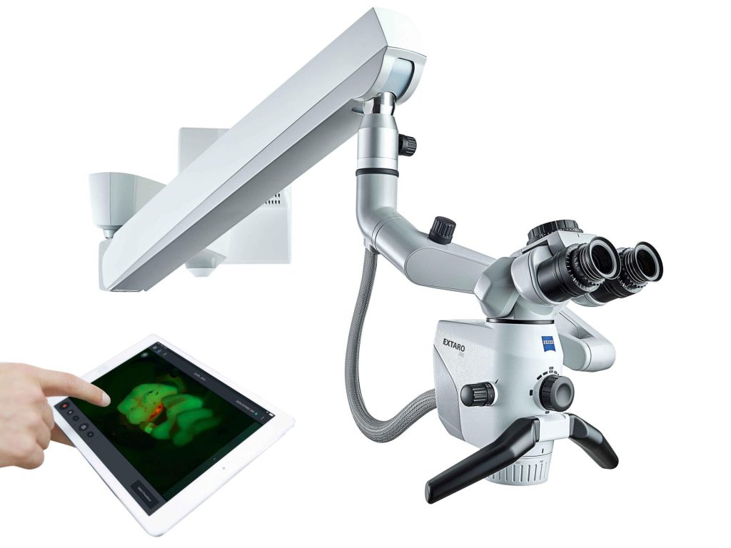
Microscope Zeiss. Extaro300
Clarity Precision Treatment effectiveness
Technology Elevating the Standards of Modern Dentistry
In modern dentistry, successful treatment does not rely solely on the experience of the dentist.
It also depends on the ability to clearly visualize the fine details of tooth structures and surrounding tissues.
The Dental Operating Microscope (DOM) has therefore become one of the key technologies that significantly enhances the quality of diagnosis and treatment. By providing high magnification and superior illumination, it enables dentists to observe extremely small details that cannot be seen with the naked eye.

The Role of the Dental Operating Microscope (DOM) in Dental Treatment
The Dental Operating Microscope (DOM) enhances magnification and image clarity in the treatment area. This allows dentists to perform procedures with greater precision and accuracy, significantly reducing the risk of potential errors.
Key benefits of the Dental Operating Microscope include:
- Improved accuracy in diagnosis
- Reduced unnecessary removal of healthy tooth structure and surrounding tissues
- More targeted and efficient treatment procedures
For these reasons, the DOM has been widely adopted across various fields of dentistry, particularly in procedures that require a high level of precision.
Applications of the Dental Operating Microscope (DOM) in Specialized Dental Procedures
Endodontic Treatment (Root Canal Therapy) The DOM allows dentists to clearly visualize complex root canal systems, including branching canals and minute internal structures within the tooth root. This enhanced visibility plays a crucial role in increasing the success rate of treatment and reducing the likelihood of retreatment.
Dental Surgical Procedures Improved visualization enables greater surgical precision, minimizing trauma to surrounding tissues and supporting better healing and recovery after treatment.
Restorative Dentistry In procedures such as fillings, crowns, and dental restorations, the DOM enhances accuracy and fit. This results in restorations that are more precise, aesthetically pleasing, and longer-lasting.
Advanced Illumination Modes with the ZEISS EXTARO 300 for Dental Procedures
The ZEISS EXTARO 300, designed specifically for dental applications, features specialized illumination modes that support different types of dental treatments and enhance clinical precision.
- True Light Mode (Orange Filter)
Provides natural white illumination that allows dentists to observe tooth color and soft tissues as close to their true appearance as possible.
This mode is ideal for diagnostic procedures and restorative dentistry. - NoGlare Mode
Reduces light reflections from restorative materials and tooth surfaces, enabling dentists to work continuously without visual disturbance or glare. - Fluorescence Mode
Helps differentiate between healthy tooth structure and areas affected by dental caries or bacterial plaque.
This improves accuracy in removing only the necessary diseased tissue while preserving healthy structures. - Green Mode
Enhances the visibility of blood vessels and soft tissues.
This mode is particularly suitable for surgical procedures and treatments that require a high level of precision.

Microscope-Assisted Root Canal Treatment
Precision That Increases the Success Rate of Treatment
Root canal treatment is a dental procedure that requires a high level of precision and attention to detail, as the internal structure of the tooth root is extremely small and complex.
The use of a Dental Operating Microscope (DOM) has become an important advancement in endodontic treatment. This technology allows dentists to clearly visualize the intricate structures within the root canal system, including calcified canals, tiny cracks, perforations, and tissues that need to be carefully removed.
How a Microscope Improves Root Canal Treatment
- The use of a dental microscope significantly enhances the quality and precision of root canal therapy in several ways:Improves accuracy in locating root canals
- Reduces the chance of missing hidden infected canals during the cleaning process: Enhances the effectiveness of root canal cleaning and disinfection
- Lowers the risk of reinfection: Minimizes unnecessary removal of healthy tooth structure, as magnified visualization allows for more precise and targeted treatment
As a result, the use of a dental operating microscope leads to higher-quality treatment outcomes and increases the likelihood of long-term success
Advantages of Using a Dental Operating Microscope (DOM) in Root Canal Treatment
- High Magnification for Enhanced Visibility
The microscope provides high magnification, allowing dentists to clearly observe the internal structures of the pulp chamber and root canal system beyond what can be seen with the naked eye. This makes it possible to detect hidden problems such as cracks, fractures, or additional root canals. - Greater Precision in Locating Root Canals
The DOM helps dentists accurately identify the openings of root canals, especially in cases where the canals are calcified, narrow, or obstructed. This ensures that treatment is performed precisely and effectively. - Preservation of Healthy Tooth Structure
With enhanced visualization, dentists can selectively remove only infected or inflamed tissue, minimizing unnecessary removal of healthy tooth structure. - Management of Complex Cases
The microscope is highly effective in treating complicated cases, such as repairing perforations in the tooth, locating separated or accessory canals, or retrieving broken instruments from within the root canal. - Higher Success Rate
Improved visibility and precision reduce treatment risks and enable more thorough cleaning of the root canal system, lowering the chances of reinfection and improving long-term outcomes. - Support for Endodontic Microsurgery
In cases of severe infection or inflammation at the root tip, the microscope assists dentists in performing precise microsurgical procedures with minimal tissue trauma.
In summary, the use of a dental operating microscope significantly enhances the effectiveness and precision of root canal treatment while helping preserve the natural tooth structure.
Contact us at Poonnakan Dental Clinic,
Dentist Consultation.
